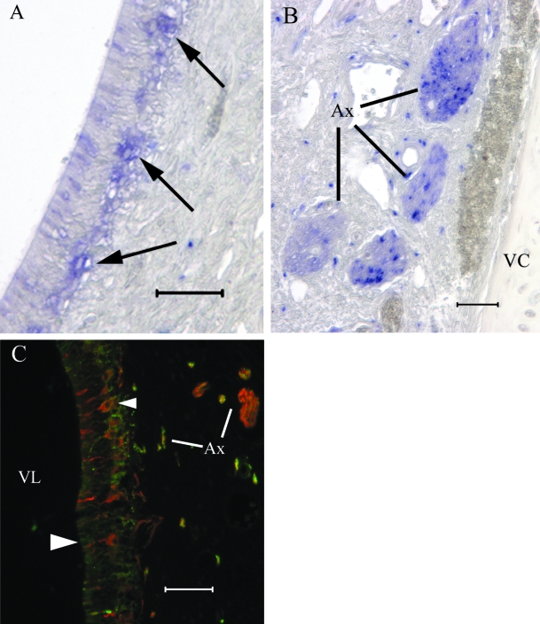
Fig. 5.
Anti-GAP43 labels clusters (A) of cell somata in the sensory epithelium (arrowheads) and VNN fascicles in the lamina propria (B). A minority of axon fascicles in the VNN nerve bundles are GAP43(+) in (B). In a preparation (C) labelled with both BT in red (Alex 594) and GAP43 in green (Alexa 488), some neurones are BT(+)/GAP43(+) (small arrowhead) and some neurones are BT(+)/GAP43(–) (large arrowhead). Some axon bundles contain both BT(+) and GAP43(+) fibres (scale bars = 50 µm).

