Fig. 3.
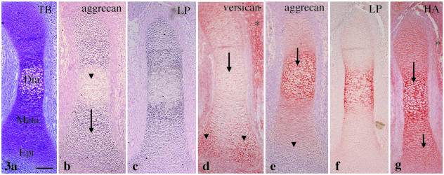
Tibial cartilage at E14. (a) Diaphysis (Dia), metaphysis (Meta) and epiphysis (Epi) are distinguishable. Toluidine blue staining. (b) Aggrecan mRNA expression is detected in the metaphysis and the epiphysis (arrow), but reduced in the diaphysis (arrowhead). (c) Link protein mRNA shows an expression pattern similar to aggrecan. (d) There is clear versican immunostaining in the mesenchyme around the cartilage (*) and in the epiphysis (arrowheads), with less toward the diaphysis (arrow). (e) There is strong aggrecan immunostaining in the diaphysis (arrow) with less toward the epiphysis (arrowhead). (f) Link protein immunostaining shows an expression pattern similar to aggrecan. (g) HA staining is detected throughout the cartilage (arrows) and mesenchyme around it (*). Width of view = 100 µm.

