Fig. 4.
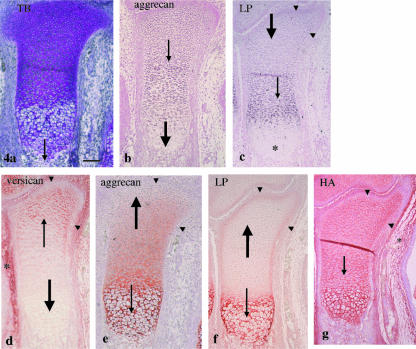
Tibial cartilage at E15. (a) Endochondral bone formation has started from the diaphysis (thin arrow). Toluidine blue staining. (b) Aggrecan mRNA is expressed in the epiphysis and metaphysis (thin arrow) but not in the diaphysis (thick arrow). (c) Link protein mRNA is strongly expressed in the metaphysis (thin arrow), weakly in the epiphysis (thick arrow) but not in the diaphysis (*). The marginal area of metaphysis and epiphysis shows relatively strong expression (arrowheads). (d) There is versican immunostaining in the mesenchyme around the tibial cartilage (*) and still in the epiphyseal end of this cartilage (thin arrow), but not in the metaphysis and diaphysis (thick arrow). (e) There is strong aggrecan immunostaining in the diaphysis (thin arrow) but there is less toward the epiphysis and none at the epiphyseal end (thick arrow). (f) There is strong link protein immunostaining in the diaphysis (thin arrow), but very weak in the metaphysis and epiphysis (thick arrow), but the marginal area of this region has moderate immunostaining (arrowheads). (g) HA staining is detected in the mesenchyme around the cartilage (*) and throughout the cartilage (thin arrow). Note that the marginal area shows no versican and aggrecan immunostainings (arrowheads in d and e, respectively) and HA staining (arrowheads in g). Width of view = 100 µm.

