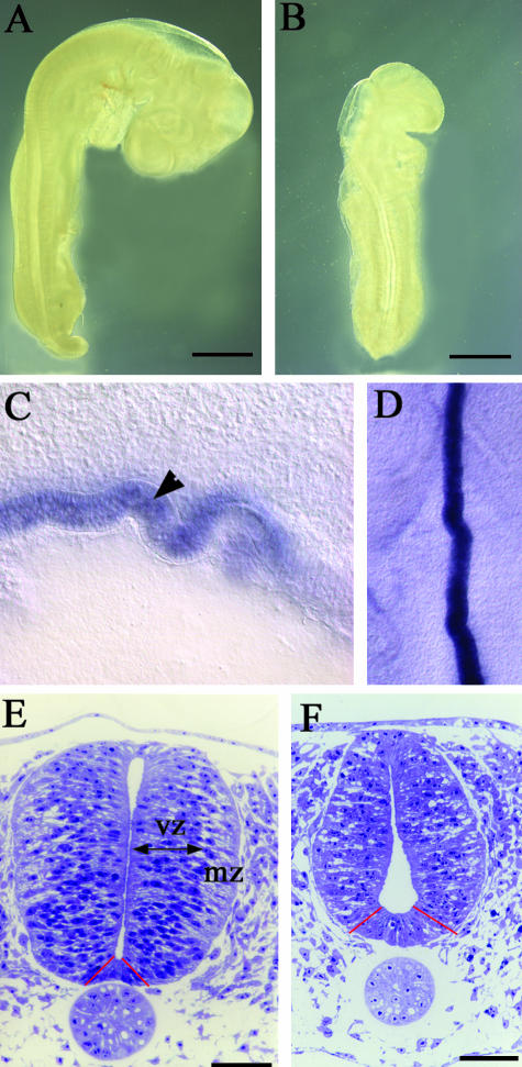
Fig. 1.
(A,B) Unstained whole embryos at stage 17 normal (A) and RA-free (B). The RA-free embryos are significantly smaller and as the posterior hindbrain cervical flexure is missing the embryos are also straighter. (C,D) Sections of shh wholemount in situ hybridizations to show the abnormal zig-zag shape of the notochord. A sagittal section is shown in C with the notochord marked with an arrowhead and a horizontal section shown in D. (E,F) Semithin (2 µm) transverse sections of stage 18 normal (E) and RA-free (F) spinal cords at the level of the forelimbs. In the normal embryo dividing cells of the ventricular zone (vz) and differentiating cells of the mantle zone (mz) are clearly distinguished and the ventral floor plate (between the red lines) is quite narrow. By contrast, the RA-free cord has many more intercellular spaces with no real distinction between the ventricular and mantle zones, the lumenal space is expanded ventrally, the floor plate is wider (between the red lines) and there is an overall decrease in size. The roof plate is also thicker (see Fig. 2). Bars in A,B = 1 mm; bars in E,F = 50 µm.

