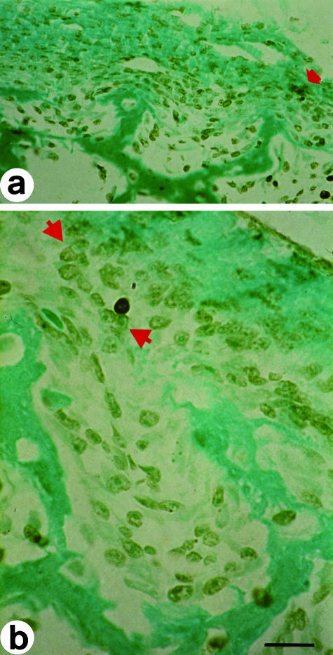
Fig. 2.
In situ end-labelling on cross-sections at the mid-diphyseal level of a newborn rabbit tibia. Labelled cells exhibit a brownish colour. (a) The arrow points to cells in apoptosis adjacent to a cord of stationary osteoblasts during SBF. (b) Cell in apoptosis behind a cord of stationary osteoblasts (between arrowsheads). LM micrographs; counterstain methyl green. Scale bars (a) = 36 µm, (b) = 15 µm.

