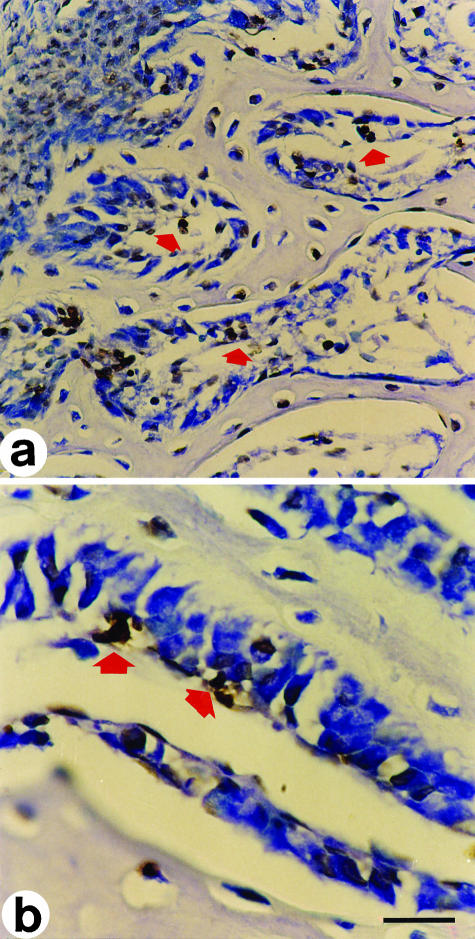
Fig. 3.
In situ end-labelling on cross-sections at the mid-diphyseal level of a 14-day-old chick embryo tibia. Labelled cells exhibit a brownish colour. (a) The arrows point to cells in apoptosis inside primitive haversian spaces, located between the movable osteoblastic laminae and the vessels, during DBF. (b) Primary haversian space lined by a moveable osteoblastic lamina; the arrows point to cells in apoptosis behind the osteoblasts. LM micrographs; counterstain toluidine blue. Scale bars (a) = 36 µm, (b) = 15 µm.

