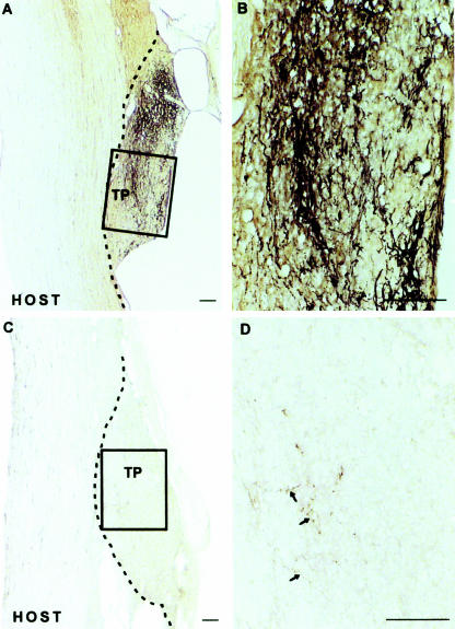
Fig. 3.
Regeneration of neonatal corticospinal axons after overhemisection lesioning, which can be seen in the untreated animals (A and B), can be completely abrogated via the addition of an inhibitor of PKA (C and D). Axons are labelled anterogradely with BDA. (Reproduced from Cai et al. 2001, with permission.)

