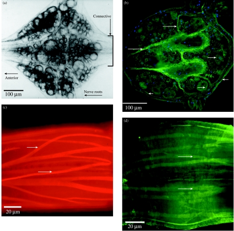
Fig. 5.
Expression of the novel peptide, designatd ReN3, in nerve cells, connective tissue and muscle of adult leech CNS. ReN3 is one of four novel clones isolated to date from the 24-h regenerating ganglion library. It has no known homologue in vertebrate genomes. (A) Isolated segmental ganglion viewed under transmitted light; ventral surface. The boxed area of the connectives is shown in C and D. (B) Horizontal section through the ganglion; orientation as in A. Immunohistochemistry using an antibody to a custom peptide shows that the encoded ReN3 peptide is widely expressed in neuron cell bodies (long arrows), in the connective tissue capsule that surrounds the ganglion (short arrows) and in the internal capsule of the ganglion (long arrows). The section is counterstained with DAPI to show nuclei. (C,D) ReN3 is also expresed in the muscle cells that extend throughout the ventral nerve cord within the connective tissue capsule. (C) A confocal section through part of the connectives that link adjacent ganglia of the ventral nerve cord (see boxed area in A), in a preparation stained with rhodamine–phalloidin to illustrate the muscle layout. (D) A confocal section through an equivalent region stained with the ReN3 antibody.

