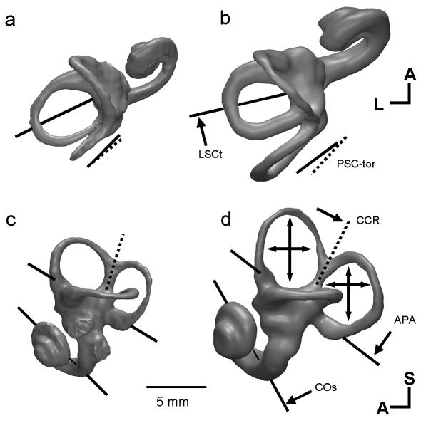
Fig. 1.
The major differences between the labyrinth of humans and great apes: CT-based 3D reconstructions, showing superior (a,b) and lateral (c,d) views of the left bony labyrinth of a female gorilla (a,c) and a modern human (b,d). In lateral view the planes of the lateral canal are aligned. Differences of orientation are indicated by single-headed arrows and differences of canal size are shown by double-headed arrows. The gorilla labyrinth is shown at 97% of its actual size to compensate for the difference in body mass (scaling follows Spoor et al. 2002b). Measurement codes as listed in Table 1 and shown in Fig. 2. A, anterior; L, lateral; S, superior.

