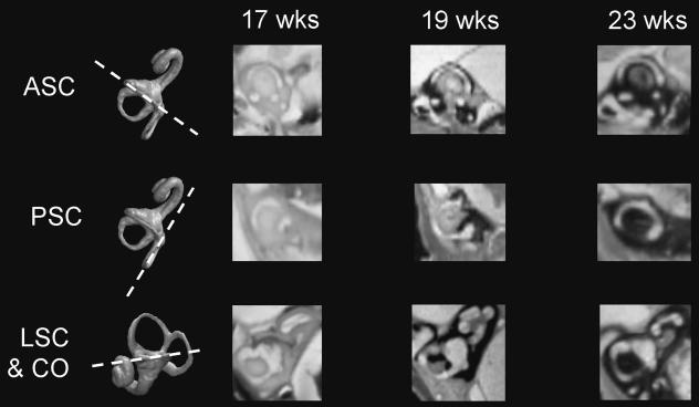
Fig. 5.
Resampled hrMRI scans showing the progress of bone formation around the: anterior semicircular canal (ASC; top row); posterior semicircular canal (PSC; middle row); lateral canal and cochlear (LSC & CO; bottom row) at 17, 19 and 23 weeks gestation. The first column shows 3D reconstructions and the plane of resampled images.

