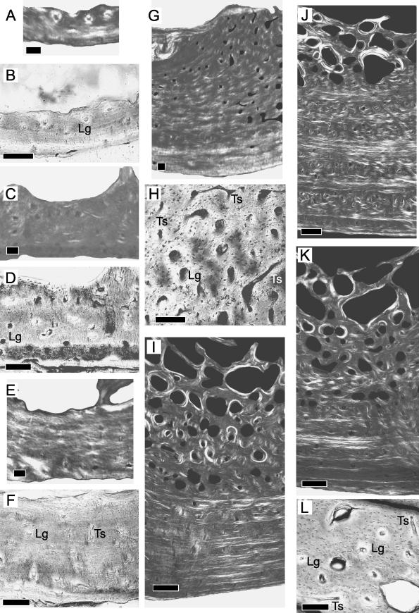
Fig. 3.
Ontogenetic sampling of the bony tissue from the ventral quadrant. Scale bars = 100 μm (A–H) and 500 μm (I–L). Superficial is down, deep is up, anterior is to the left, posterior is to the right. Specimens 1–7, general view under circularly polarized light (A,C,E,G,I,J,K). Detailed view under non-polarized light (B,D,F,H,L). Longitudinal canal (Lg), transverse canal (Ts).

