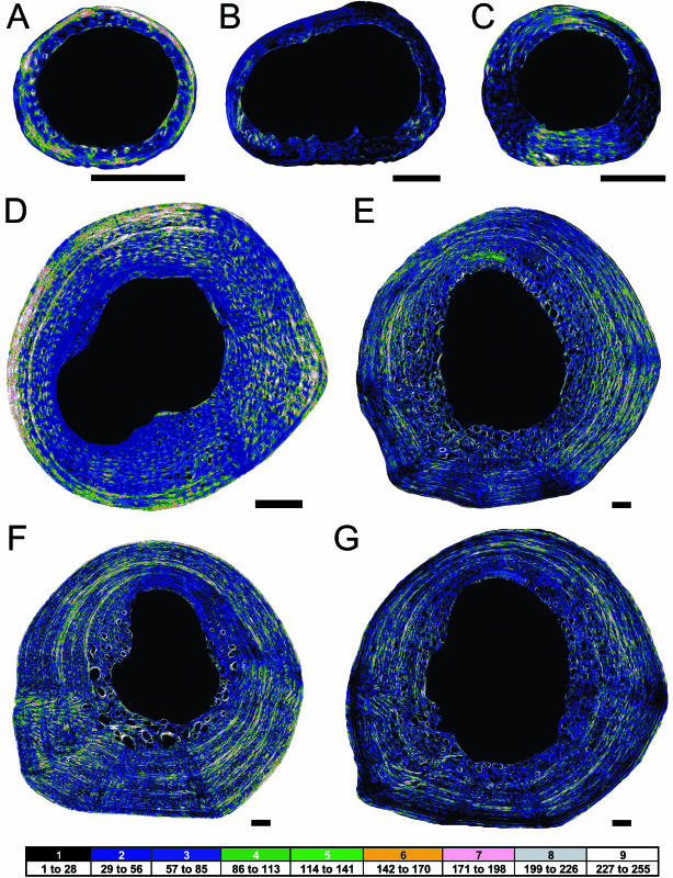
Fig. 4.
Ontogenetic series of collagen fibre distribution of the alligator femur. Scale bar = 1 mm. Specimen 1 (A), specimen 2 (B), specimen 3 (C), specimen 4 (D), specimen 5 (E), specimen 6 (F) and specimen 7 (G). Darker colours represent more longitudinal collagen fibres, and brighter colours represent more transverse collagen fibres. Medullary and vascular spaces are black. Dorsal is up, and anterior is left.

