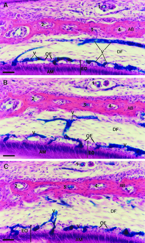
Fig. 3.
Light micrographs of three serial sections (20 μm thick each) of the same lateral region of the enamel organ of a molar tooth germ from a 5-day-old rat injected with blue Indian ink. Blood vessels in the dental follicle (DF) and in the enamel organ (EO) are filled with blue Indian ink. The enamel organ (EO) shows polarized ameloblasts (AM) at the stage of enamel formation and involuting stellate reticulum. These consecutive sections were outlined for a 3D computer reconstruction of the blood vessels (V), as shown in Fig. 4. Structures used as fiducial points for alignment of the three serial sections are indicated by the numbers 1, 2, 3 and 4. AB, alveolar bone; OE, outer enamel epithelium. H&E. Scale bar = 40 μm.

