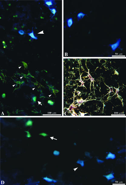
Fig. 7.
Multiple labelling experiments. Three neuroanatomical tracers were injected into different muscles of the same side of the face. A, photomicrograph obtained by simultaneous dark-field illumination and epifluorescence. Large arrowhead, retrogradely labelled neuron after injection of Fast blue (FB) into the LELSAN; small arrowhead, retrogradely labelled neurons after injection of CTB-HRP into the auricularis superior; arrow, retrogradely labelled neuron after injection of Diamidino yellow (DY) into the frontalis. B, epifluorescence photomicrograph showing retrogradely labelled neurons after injection of FB into the LELSAN. C, dark-field photomicrograph showing retrogradely labelled neurons and their dendritic arborization after injection of CTB-HRP into the orbicularis oculi (superior portion). D, epifluorescence photomicrograph showing retrogradely labelled neurons after injection of DY (arrow) into the zygomaticus and of FB (arrowhead) into the orbicularis oris (inferior portion).

