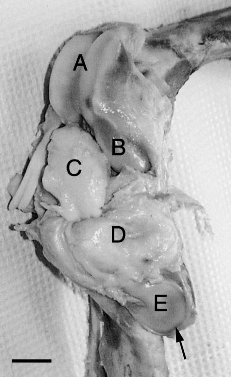
Fig. 1.
Right stifle joint tissues of swine showing articular cartilages of trochlea (A), medial femoral condyle (B) and patella (E), and the inner tissue (C) separated from the outer tissue (D). The inner and outer tissues show the ventral and dorsal surfaces, respectively. An arrow shows the proximal end of the patella. Scale bar = 2 cm.

