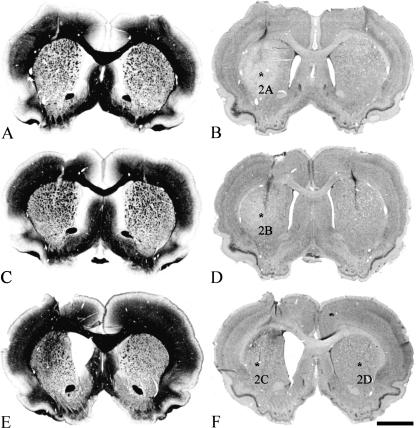
Fig. 1.
Histological evaluation of successful QA lesions of the CPu. Frontal sections through the injection site 7 days (A,B), 14 days (C,D) and 29 days (E,F) after a unilateral QA lesion (left hemisphere) are illustrated. Myelin stainings are shown in the left column (A,C,E) whereas Nissl stainings are shown to the right (B,D,F). The decreasing tissue volume of the CPu and the enlargement of the lateral ventricle over time after lesion, compared with the sham-lesioned side, are clearly visible. Asterisks (B,D,F) indicate the corresponding regions shown in Fig. 2. Scale bars, 500 μm (A–F).

