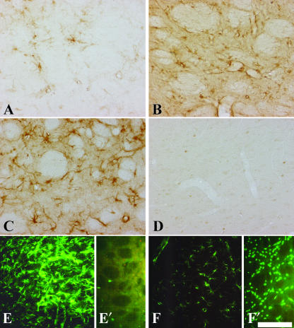
Fig. 3.
Immunohistochemical detection of CNTF, GFAP and NeuN in the QA-lesioned and sham-lesioned contralateral CPu. Increasing staining intensity for CNTF 7 days (A), 14 days (B) and 29 days (C) after a QA lesion is shown. The sham-lesioned CPu (D) does not show any immunoreactivity for CNTF. In the QA-lesioned CPu 29 days post-lesion (E), GFAP immunoreactivity is strongly increased compared with sham-lesioned CPu (F). Twenty-nine days after QA lesion the loss of neurons is clearly detectable by immunostaining against the neuronal nuclei antigen in the lesioned CPu (E′), compared with the sham-lesioned (F′) CPu with many NeuN-immunoreactive cells. Scale bars, 100 μm (A–D), 175 μm (E,F).

