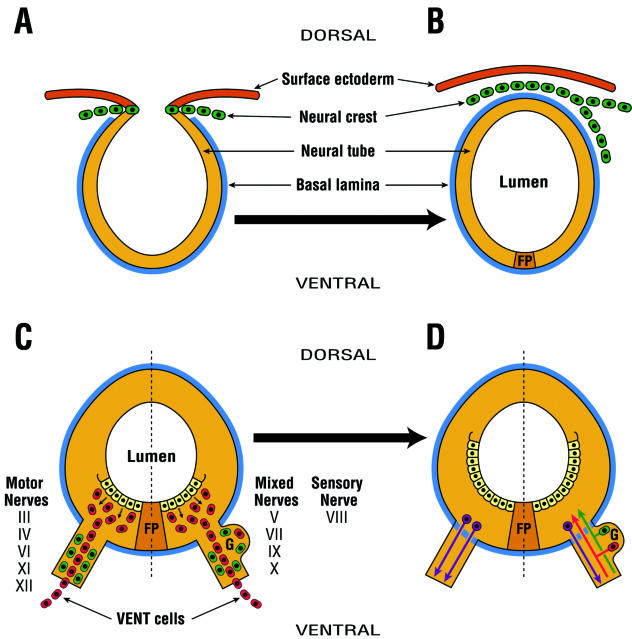
Fig. 1.
The concept of emigration of two separate and distinct neural tube-derived cell populations: neural crest and VENT cells. (A) Neural crest cells (green) arise from the dorsal region of the developing neural tube. They are HNK-1+. (B) Neural crest cells emigrate from the dorsal midline, and contribute to the development of the PNS and a variety of neural and non-neural tissues. This occurs in the cranial neural tube of the chick embryo between HH stages 9 and 11 (about E2). (C) VENT cells (red) arise from the ventral region of the neural tube. They are HNK-1−. VENT cells emigrate at the exit/entry site of all cranial nerves of the midbrain and hindbrain through a transient breach in the basal lamina (blue) surrounding the neural tube. In the chick, this begins to occur at about HH stage 16 (E3), i.e. considerably after the emigration of neural crest, but before the outgrowth/ingrowth of axons from the neural tube to the nerves. Prior to the emigration of VENT cells, the cranial nerves contain neural crest and/or placodally derived HNK-1+ cells. After this emigration, the nerves also contain HNK-1− VENT cells. Most VENT cells continue their migration to target tissues of these cranial nerves. (D) About 1 day later (i.e. E4), the transient breach is sealed. By E5, some of the VENT cells (red) that remain in the ganglia, and neural crest and/or placodally derived cells (green), differentiate into sensory neurons, with axons entering the neural tube through a fenestrated basal lamina. Motor neuron (purple) axons similarly exit the neural tube. FP, floor plate.

