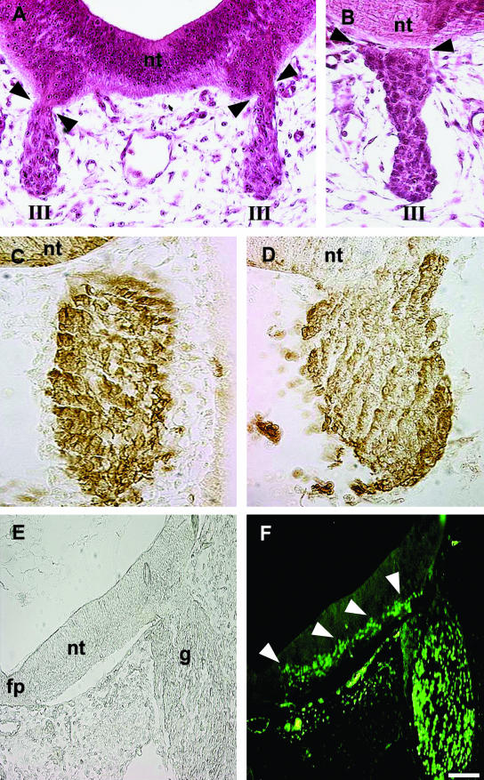
Fig. 2.
Circumstantial evidence for the VENT cells from normal (i.e. experimentally unmanipulated) embryos. The ventral neural tube is directly connected transiently with the cranial nerves. (A,B) H&E-stained cross-sections of quail embryos. nt, midbrain neural tube; III, the third cranial nerve. Continuity between the ventral neural tube and the nerve is seen at E3 (A, arrowheads) but not at E4 (B, arrowheads). Differential distribution of the HNK-1 antigen. (C,D) Sections through the hindbrain neural tube and fifth cranial nerve of quail embryos at E2.5 (C) and E3 (D), immunostained with anti-HNK-1 antibody. HNK-1+ cells are dark brown, whereas HNK-1− cells are unstained. nt, neural tube. Prior to continuity with the neural tube, the ganglion comprises HNK-1+ cells (C). After continuity is established at E3, the ganglion also contains regions of HNK-1− cells, continuous with the neural tube (D). The HNK-1− cells are mainly in the central regions, whereas the HNK-1+ cells are mainly in the periphery of the ganglion. (E,F) Expression pattern of Islet-1. A section through the hindbrain neural tube and fifth cranial nerve of an E3.5 chick embryo is shown in bright-field (E) and fluorescence (F) after immunostaining with anti-Islet-1 antibody. fp, floor plate; g, ganglion; nt, neural tube. Arrowheads point to a trail of Islet-1+ cells in the ventral neural tube, extending into the ganglion. Scale bar, (A) 32 µm; (B) 28 µm; (C) 32 µm; (D) 29 µm; (E, F) 75 µm.

