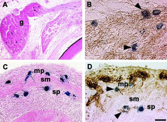
Fig. 7.
Differentiation of VENT cells into nerve tissue. Emigration of VENT cells into nerve tissue was monitored by tagging ventral neural tube cells with lacZ retrovirus at stage 14. Labelled cells were subsequently detected histochemically (blue-stained cells, A–D). (A)Some of the VENT cells that had emigrated into the trigeminal ganglion (g) remained in the ganglion by E6. (B)Some of these VENT cells expressed β-tubulin (arrowheads), a marker for neuronal tissue (brown immunostained cells). (C)A section of an E9 duodenum is shown. Some VENT cells migrating in association with the vagus nerve populated the region of the ENS in the gut. mp, myenteric plexus; sp, submucosal plexus; sm, smooth muscle layer. (D)Some of these VENT cells expressed β-tubulin, indicating differentiation into neurons of the ENS (arrowheads). Scale bar, (A) 119 µm; (B) 14 µm; (C) 21 µm; (D) 15 µm.

