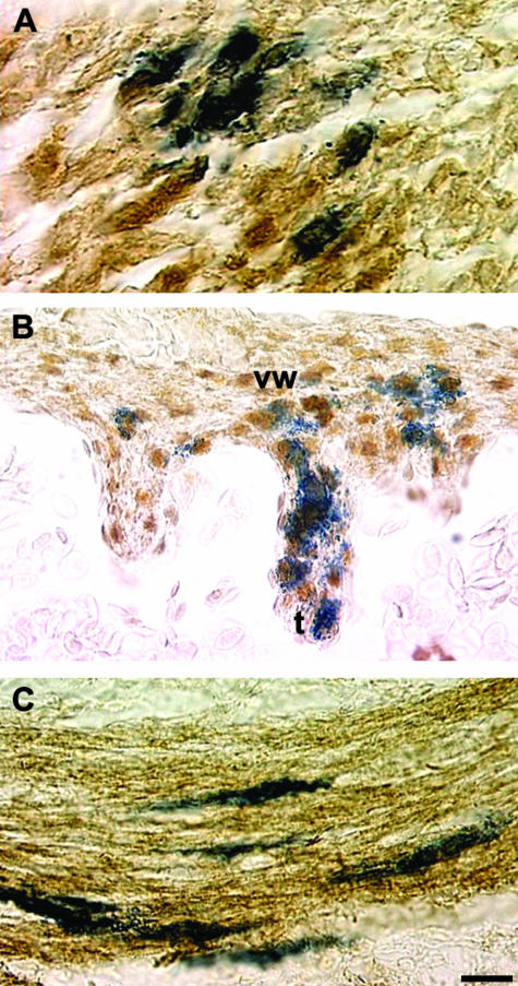
Fig. 8.
Differentiation of VENT cells into muscle tissue. Pre-emigratory VENT cells were labelled by injection of LacZ-expressing retrovirus into the lumen of the neural tube of stage 14 chick embryos. Emigrated, labelled cells in tissues were subsequently detected histochemically (blue-stained cells, A–C). (A)By E7, some lacZ-labelled VENT cells have populated craniofacial muscle tissue, and are immunopositive for skeletal muscle fast myosin (brown colour), indicating differentiation into skeletal muscle. (B)By E8, some VENT cells in cardiac muscle are immunopositive for α-actin smooth muscle, which is also expressed in heart tissue, indicating differentiation into cardiac muscle. vw, ventricular wall; t, trabecula. (C)By E11, some VENT cells in the circular smooth muscle layer of the duodenum express α-actin smooth muscle, demonstrating differentiation of VENT cells into smooth muscle cells. Scale bar, (A) 12 µm; (B) 17 µm; (C) 13 µm.

