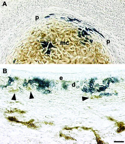
Fig. 9.
Differentiation of VENT cells into connective tissue. Pre-emigratory VENT cells were labelled by injection of LacZ-expressing retrovirus into lumen of the midbrain or hindbrain neural tube of stage 14 (E3) chick embryos. Emigrated, labelled cells in tissues were subsequently detected histochemically (blue-stained cells, A and B). (A) By E7, VENT cells were present in Meckel's cartilage (mc) and the surrounding perichondrium (p). HNK-1+ cells (brown immunostain) are absent in the perichondrium but are prominent in the cartilage. The VENT cells are HNK-1−. (B)VENT cells populate the dermal layers (d) of the craniofacial skin by E7. Unlike other cells in the dermis (arrowheads, brown immunostain), they are HNK-1−. e, epidermis. Scale bar, (A) 22 µm; (B) 13 µm.

