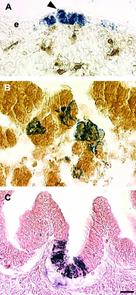
Fig. 10.
Differentiation of VENT cells into epithelium. Pre-emigratory VENT cells were labelled by injection of LacZ-expressing retrovirus into lumen of the neural tube of stage 14 (E3) chick embryos. (A)VENT cells populate the epidermal layers of the craniofacial skin by E7. Labelled VENT cells (blue) are observed in all layers of the epidermis (e). In some cases columns of labelled cells rise from the basal layers to the superficial layers (arrowhead). Brown immunostained cells in the dermis are HNK-1+, unlike the VENT cells. (B)By E11, many VENT cells in the liver express albumin (brown immunostain), a marker for hepatocytes. (C)Some VENT cells in the duodenum differentiate into epithelial cells. The VENT cells are often located in the deeper layers of the villi, which is where the proliferating stem cells are located. Scale bar, (A) 13 µm; (B) 8 µm; (C) 13 µm.

