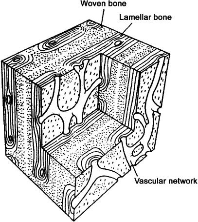
Fig. 2.
Block diagram of the general histological type of primary bone known as ‘laminar’, ‘plexiform’ or ‘fibrolamellar’ (Currey, 2002). This diagram shows representative primary vascular canal types. The distance between the vascular networks is 100–200 µm in bones from different species. It should be noted that the more obvious circularly orientated canals of highly orientated laminar tissue (i.e. ‘laminar vascularization’ patterns) are not depicted in this diagram. See Fig. 1 and text for further discussion of the distinctive vascular patterns that can be found in ‘laminar’ bone. (Adapted from Zioupos & Currey (1994, p. 983) with permission of Kluwer Academic Publishing.)

