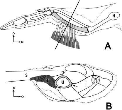
Fig. 3.
Dorsal-to-ventral view (A) and cross-section view (B) of left avian wing: H, humerus; U, ulna (shaded); R, radius; Cr, cranial; D, dorsal; M, medial; C, secondary covert feather; T, ‘tether’ of secondary feather (S) sheath (sheath = calamus wall; calamus = quill). The lower drawing is a proximal-to-distal view of the left forelimb transversely sectioned at mid-diaphysis. A secondary feather is shown diagrammatically in profile; the remainder of the drawing represents the actual cross-section. There are 18 secondary feathers in domesticated turkeys, and all of these ‘attach’ via ‘tethers’ along the caudal ulna (Lucas & Stettenheim, 1972). The arrow indicates the location of a firm insertion of muscles into the cranial cortex. (The upper figure is modified after Figs 18,4 and 18,8 in the Guild Handbook of Scientific Illustrations (Hodges, 1989) with permission of the artist Nancy Halliday and the publisher.)

