Fig. 1.
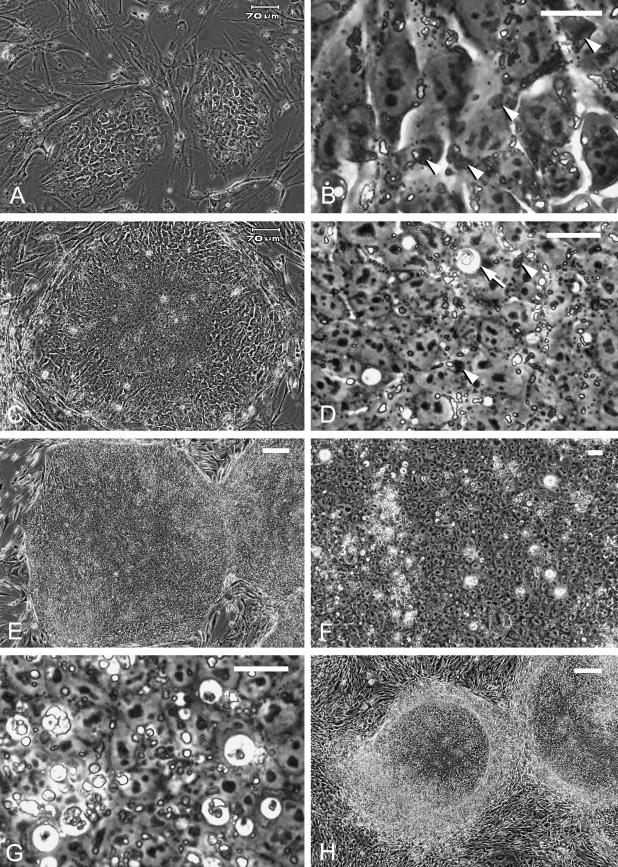
Images of living human ES cell colonies under phase contrast microscopy. (A) Initial-stage colonies 24 h after splitting. These colonies have a mosaic appearance with discernible cell–cell borders. Bar = 70 µm. (B) High magnification of one of the colonies in A. Note the dark deposits (arrowheads) and bright granules in the cytoplasm. Bar = 20 µm. (C) A colony 4 days after splitting. Central compaction and peripheral mosaic appearance can be seen. Bar = 70 µm. (D) High magnification of the colony in C. Dark deposits (arrowheads) and fine, bright granules are observed in the cytoplasm of the compact cells. A coarse particle with a halo, presumably a phagosome, can be seen (arrow). Bar = 20 µm. (E) Large, undifferentiated colonies 7 days after splitting. The colony in the centre is about 1.5 mm in diameter. Bar = 200 µm. (F) Coarse particles are dispersed heterogeneously in a large colony 7 days after splitting. Bar = 20 µm. (G) Coarse particles with or without a halo 5 days after splitting; some presumably represent apoptotic cells/bodies. Bar = 20 µm. (H) Spontaneously differentiated colonies 6 days after splitting show a peripheral differentiated area. Bar = 200 µm.

