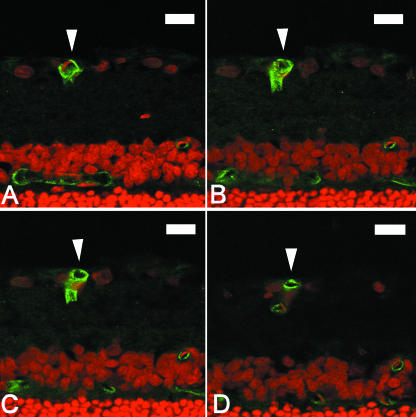
Fig. 2.
(A–D) Four serial optical sections obtained by confocal microscopy from the same 20-µm-thick frozen section of retina. Each optical section is 2 µm thick and each image is separated by a distance of 2 µm. Cell nuclei are stained with propidium iodide (red). Blood vessels are stained with biotinylated griffonia simplicifolia isolectin B4 and labelled with FITC (green). A typical blood vessel branch point is demonstrated. In (A) a single blood vessel is visible in the retinal ganglion cell layer (indicated by arrow). In (B) and (C) the vessel is seen to branch to form two individual vessels sharing a common wall. In (D) the vessel has completely separated to form two distinct vessels. Scale bar = 20 µm.

