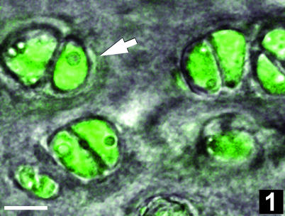
Fig. 1.
Fluorescence photomicrograph of a tangential section through the superficial zone of rabbit femoral condyle articular cartilage. Note the walnut-in-the-shell appearance of chondrocyte pairs: chondrocytes exhibit green fluorescence with the vital dye calcein-AM; the pericellular matrix delimiting the chondron (arrow) is visible in the transmitted light channel of the confocal microscope (composite fluorescence–differential interference contrast image). Scale bar, 10 µm.

