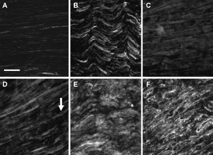
Fig. 1.
Immunofluorescent staining of NCAM (A–C) and N-cadherin (D–F) in normal rat sciatic nerve (A,D), and proximal (B,E) and distal stumps (C,F) after axotomy. Monochrome images were taken for quantification in order to minimize loss of signal intensity. Images were taken ∼2 mm proximal or distal to the injury site. Low levels of staining are seen in all normal nerve sections (A,D). N-cadherin stains structures that resemble Schwann cells with slender processes originating from a more densely stained cell body (arrow). Fifteen days after axotomy there appears to be an increase in staining intensity for NCAM (B) and N-cadherin (E). A similar increase in expression is seen in distal stumps for N-cadherin (F), although NCAM levels do not appear to stain as intensely (C). Scale bar, 50 µm (objective × 40). NB. Increased exposure times were used for normal nerve sections to allow staining to be adequately visualized.

