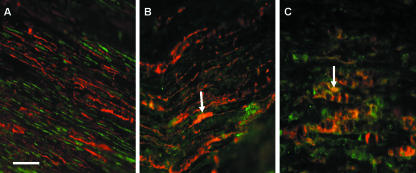
Fig. 2.
Immunofluorescent double-staining with S-100 (red fluorescence) and either NCAM (A), N-cadherin (B) in normal rat sciatic nerve or N-cadherin in the distal stump after axotomy (C) (all adhesion molecules show green fluorescence). NCAM staining in normal nerve sections does not appear to co-localize with S-100, unlike N-cadherin (arrow in B) (orange/yellow staining areas). In the distal stump after axotomy N-cadherin staining co-localizes with S-100 (arrow in C), indicating that Schwann cells may be responsible for the high levels of expression of N-cadherin in distal stumps. Scale bar, 50 µm (objective × 40). NB. Increased exposure times were used for normal nerve sections to allow staining to be adequately visualized.

