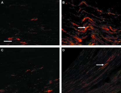
Figure. 3.
Immunofluorescent double-staining with PGP9.5 (red fluorescence) and NCAM (green fluorescence). In the centre of the crushed zone after 2 days (A), PGP9.5 expression is minimal and undistinguishable from PGP9.5 staining in uninjured nerve due to the physical destruction caused by nerve crush. However, after 7 days (B) PGP9.5-positive staining is seen corresponding to regenerating axons co-localizing with NCAM staining (arrow) (orange/yellow staining areas). In the section of nerve distal to the crush injury after 15 days (C) there is little PGP9.5 staining, indicating regenerating axons may not have crossed the crushed zone; however, in the same region after 30 days (D) some regenerating axons appear to be present. Again NCAM appears to be expressed in co-localization with PGP9.5 in the distal zone 30 days after crush (arrow in D) (orange/yellow staining areas). Scale bar, 50 µm (objective × 40).

