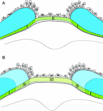
Fig. 7.
Schematic illustration of the intervertebral region at two developmental stages, demonstrating the possible mode of growth. Layer 2 is shown in different shades of green, and the vertebral part of this layer indicated by vertical lines, which is covered by layer 3, is mineralized. Layer 3 is shown in shades of blue. Osteoblasts and fibroblasts are indicated. (A) Layer 2 forms a continuous collagenous structure, which is deposited by fibroblasts in the intervertebral region. The layer consists of a mineralized (I) and a non-mineralized (II) part. (B) Later stage where the part of layer 2, which formed the intervertebral ligament (II) in A, is covered by layer 3 (light blue), and is subsequently mineralized. During this process, new non-mineralized collagen is deposited in the intervertebral region (III).

