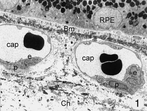
Fig. 1.
Electron microscopy of the choriocapillaris: a thin layer of fenestrated endothelial cells lie on a thin basement membrane. Only very rarely pericytes can be seen. Magnification ×8000. (Eye removed due to neovascular glaucoma.) Abbreviations for this and other figures: Bm, Bruch's membrane; RPE, retinal pigment epithelium; Ch, choroid; m, mitochondria; Gc, special cell; cap, capillary; e, endothelial cell; er, ergastoplasmatic reticulum; p, pericyte; d, cellular process.

