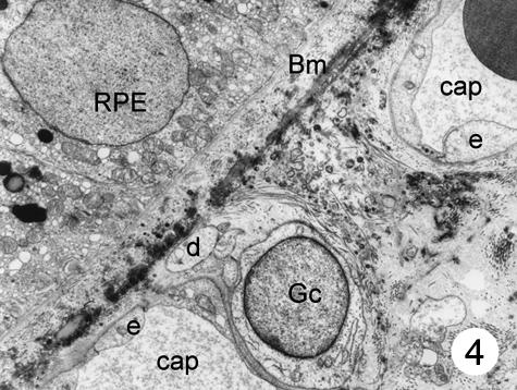
Fig. 4.
The cell body and some processes of a special cell can be seen in the intercapillary connective tissue, next to a choriocapillary. One of the processes reaches the elastic layer of Bruch's membrane. No pericytes can be seen. Magnification ×8000. (Eye removed due to intraocular melanoma.) For abbreviations, see legend to Fig. 1.

