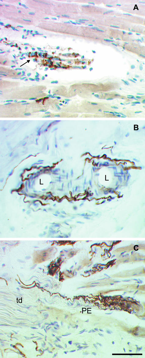
Fig. 2.
Photomicrographs of neural structures in longitudinal sections of sheep and human extraocular eye muscles labelled with SNAP-25 antibodies and counterstained with hemalaun. (A) Identified muscle spindle in a sheep inferior rectus muscle. With SNAP-25 immunolabelling, a nerve fibre which enters the collagenous capsule is visualized. On the intrafusal muscle fibres an annulospiral ending which winds around the fibres is clearly SNAP-25 positive (arrow). (B) Blood vessels in the tendon of a human extraocular muscle. Using SNAP-25 antibodies, a fine network of varicose nerve fibres surrounding the lumen (L) of the vessels could be identified. (C) A palisade ending at the myotendinous junction of a human extraocular eye muscle. SNAP-25 antibodies completely label the palisade ending (PE), the axon which enters from the tendon (td) as well as the bunch of branches and small terminals which terminate on a muscle fibre. Scale bar, 50 μm.

