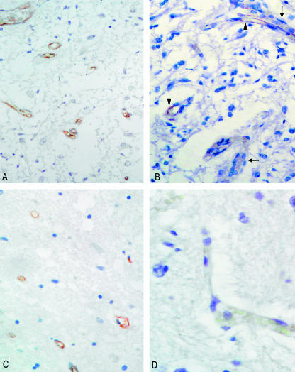
Fig. 3.
(A) Section of area postrema of an adult subject, stained with anti-von Willebrand factor, showing positive reaction of all vessels (×20). (B) Area postrema of an infant subject, showing coexistence of positive (arrowheads) and negative (arrows) vessels (×25). (C) Section of solitary tract nucleus of adult, with positive staining of all vascular structures (anti-von Willebrand factor, ×25). (D) Vessel in solitary tract nucleus of infant, showing negative staining at immunohistochemistry (anti-von Willebrand factor, ×40).

