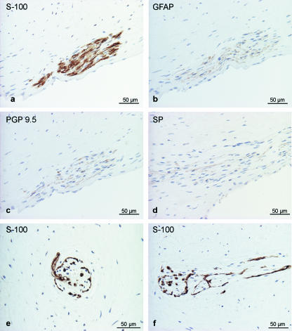
Fig. 3.
Nerve fibres in the tusk pulp detected by immunohistochemical staining for general neural markers. (a) S-100, (b) GFAP, (c) PGP 9.5, (d) SP-positive axons within the nerve bundle. Special nerve fibre terminals resembling Ruffini endings were found within the pulp (e, cross-section; f, longitudinal section) stained for S-100 protein.

