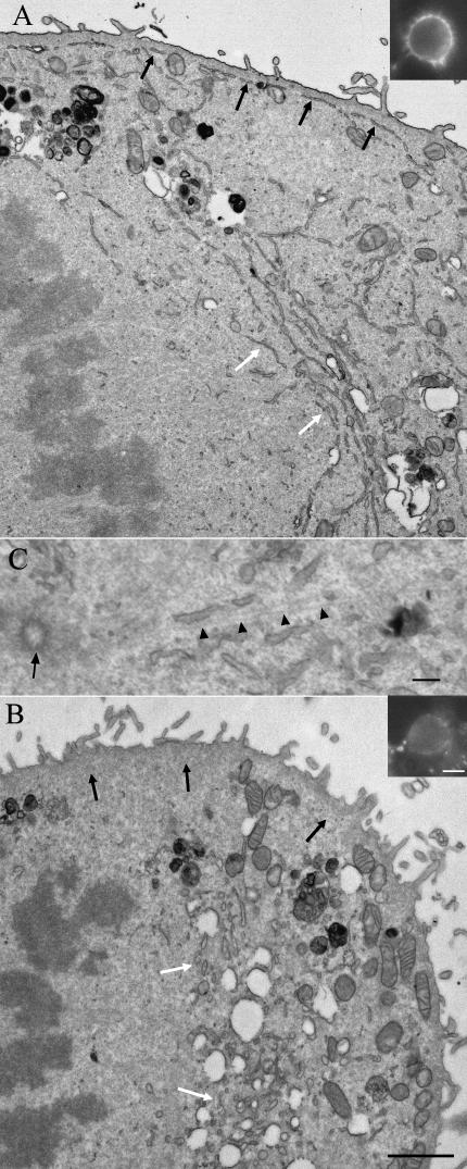
Fig. 5.
Endoplasmic reticulum in thin sections of epoxy-embedded metaphase monolayer HeLa cells. The peripheral RER in untreated cells (A) was found at a constant distance from the plasma membrane and closely applied to the cortex (black arrows), which was shown to contain actin using fluorescently labelled phalloidin (inset). Deeper in the cell and close to the spindle there is a concentration of parallel RER cisternae (white arrows). In cells treated for 2 h with 5 µm Latrunculin A (B) peripheral RER is no longer present (region of the black arrows) but the RER is visible in the juxta-spindle region and is now present as shorter, more rounded section profiles (region of the white arrows). The peripheral actin staining is now very weak (inset to B). Mitochondria (labelled M in A and B) become concentrated in the periphery after Latrunculin A treatment. (C) Shows close association of ER cisternae with microtubules (arrow heads) radiating from the centriole (arrow). Scale bars: A and B, 2 µm; insets, 10 µm; C, 150 nm.

