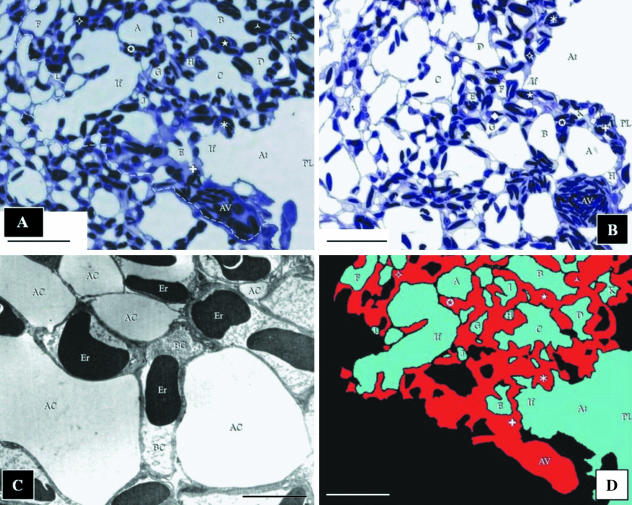
Fig. 1.
(A) The first of the 71 images of the sections of the exchange tissue of lung of the muscovy duck, Cairina moschata, used for the reconstruction. The area that was reconstructed is located to the right of the dashed line. The parabronchial lumen (PL) gives rise to an atrium (At), which narrows to form an infundibulum (If). The surrounding parenchyma (exchange tissue) comprises ACs of variable sizes (letters) and BCs (symbols). The BCs drain into the atrial vein (AV). Scale bar, 25 μm. (B) The last section used in the reconstruction. The atrial vein (AV) appears larger in diameter than in the first section (A). This indicates that blood flows in the direction from the first to the last section, draining the BCs that lie along its length. The parabronchial lumen (PL) can be seen giving rise to an atrium (At), and an infundibulum (If). ACs are labelled using letters and BCs using symbols. Scale bar, 25 µm. (C) Transmission electron micrograph prepared to establish that for the purposes of the present study (3D reconstruction), the fixation of the lung was adequate. AC, air capillaries; BC, blood capillaries containing erythrocytes (Er). Scale bar, 5 μm. (D) The reconstructed area shown in A (at the same magnification, orientation, and with the structural components labelled identically) indicates how the air- and the vascular elements were identified and colour coded. The areas in black comprised: (a) ACs (in the construction) of which the continuity to the parabronchial lumen could not be established, (b) of parts (outside of the reconstruction) that fell outside of the limit of the reconstructed part, and (c) the tissue barrier and the BCs not containing erythrocytes and blood plasma. These regions were not included in the reconstruction. Colour coding: blue, air spaces; red, blood. Scale bar, 25 μm.

