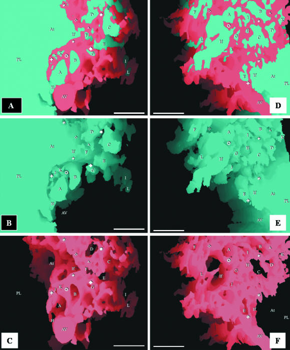
Fig. 3.
Respective views of combined, airway and vascular reconstructions. The labelling is identical in each of the three sequential figures (A–C and D–F) to show the topographical locations of the same structures. The parabronchial lumen (PL) can be seen giving rise to an atrium (At), which narrows to form an infundibulum (If). The exchange tissue (parenchyma) is composed of ACs (letters) and BCs (symbols). The BCs can be seen draining into an atrial vein (AV). All scale bars, 25 μm.

