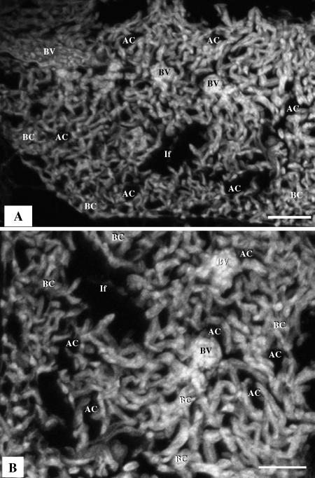
Fig. 8.
(A) Three-dimensional reconstruction based on 17 serial sections generated by confocal laser scanning microscopy (CLSM), showing blood vessels (BV), air capillaries (AC), tubular, interconnected blood capillaries (BC) and infundibulae (If). Scale bars 50 µm. (B) A single optical section produced by CLSM; the BCs are sausage-like in shape while the ACs (air spaces) are saccular: the gas exchange units are not mirror images.

