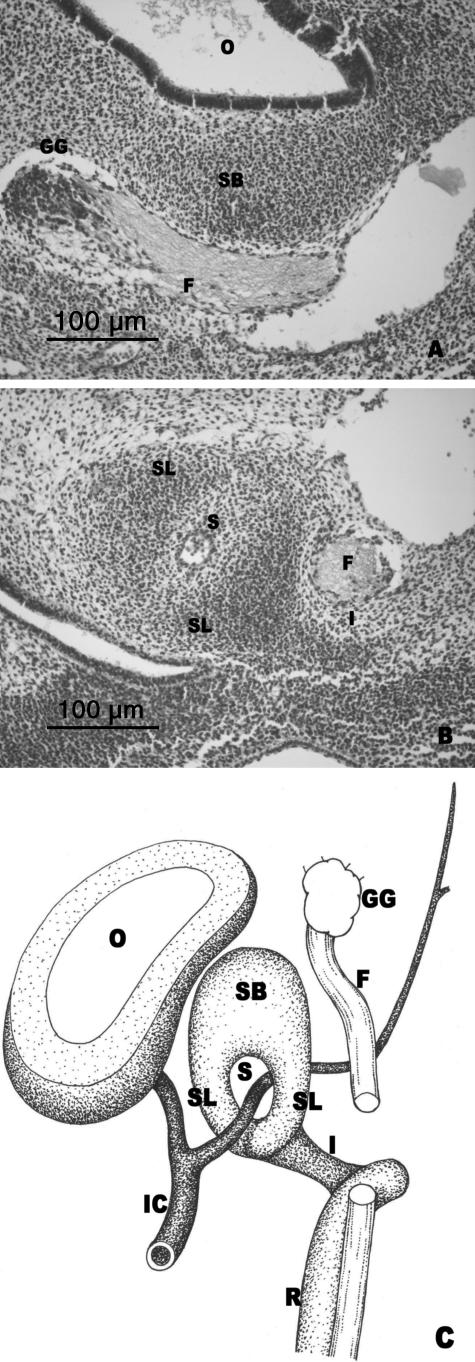
Fig. 4.
(A,B) Human embryo GV-6 (13 mm CR; O'Rahilly's stage 17). Transverse sections. (C) Diagrammatic representation of the stapedial anlage. Superior portion of the stapedial anlage (SB) medial to the facial nerve (F). Geniculate ganglion (GG). Otic vesicle (O). Anlage of the stapedial limbs (SL) separated by the stapedial artery (S). The interhyale (I) connected with the stapedial limb (SL). Facial nerve (F). Internal carotid artery (IC). Reichert's cartilage anlage (R).

