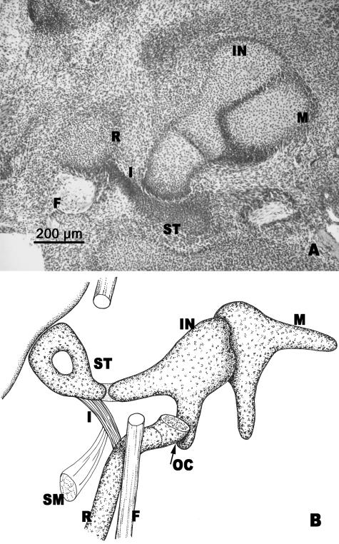
Fig. 6.
(A) Human embryo AR (20.5 mm CR; O'Rahilly's stage 20). Frontal section. (B) Diagrammatic sketch of the ossicles of the middle ear. Malleus (M). Incus (IN). The interhyale (I) lies between the stapes (ST) and the cranial portion of Reichert's cartilage (R). Facial nerve (F). Otic capsule (OC). Stapedial muscle (SM).

