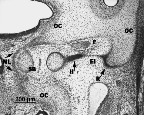
Fig. 8.
Human embryo BR-4 (28 mm CR; O'Rahilly's stage 23). Frontal section. The stapedial base (SB) is adjacent to the mesenchymal lamina (ML) that closes the anlage of the oval window in the otic capsule (OC). The internal segment of the interhyale (II) that forms the tendon of the stapedial muscle is clearly differentiated from the very thin external segment (EI) that reaches Reichert's cartilage (R) and, in turn, is joined to the otic capsule (OC).

