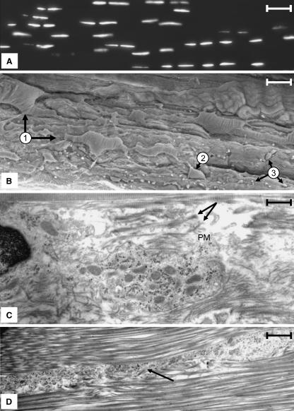
Fig. 1.
(A) Frozen section of a rabbit MCL stained with DAPI for DNA. Nuclei are arranged in rows that pass along the long axis of the tissue (right–left). Note the rows cannot be followed continuously for long distances. Scale bar = 40 µm. (B)SEM micrograph of a rabbit MCL that has been torn along the long axis of the tissue revealing rows of cells (arrow 1). The cells are large (in many instances a portion of the cell is buried in the collagen matrix; compare the two cells denoted at 1), having a complex morphology and displaying prominent cytoplasmic extensions. Portions of these extensions permeating the collagen matrix are denoted at 3. The diversion of a row into another plane within the tissue is shown at 2. Scale bar = 25 µm. (C) A region between two adjacent cells within a row illustrating the pericellular matrix, which is bordered by large collagen fibres characteristic of the ECM. The pericellular matrix is populated, with collagen fibres of varying sizes (arrows) and distribution paths (arrow) as well as abundant vesicles. Scale bar = 11 µm. (D) A vesicle-filled seam representing an extension of the pericellular matrix that extends between collagen bundles into the ECM. These seams contain cytoplasmic extensions of the ligament cells as well as vesicular material similar that that seen in the pericellular matrix adjacent to the cell. Scale bar = 0.5 µm.

