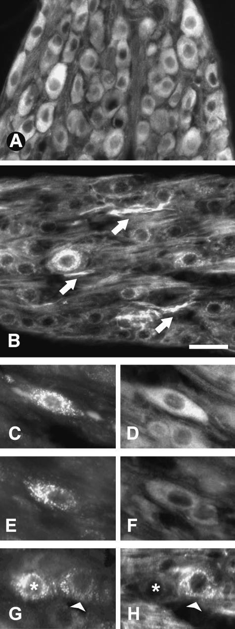
Fig. 1.
Cryostat tissue sections of rat thoraco-lumbar sympathetic chain ganglia showing neurons immunostained for TrkA (A) and p75 (B). Arrows in B indicate p75-positive nerve bundles. (C–E) Fluorogold-traced (FG) uterine-projecting sympathetic neurons displaying, respectively, high (D) and low (F) levels of TrkA. Fluorogold-labelled neurons shown in G (asterisk and arrowhead) also show marked differences in their levels of p75 (H). Scale bars: A,B = 40µm; C–H = 25µm.

