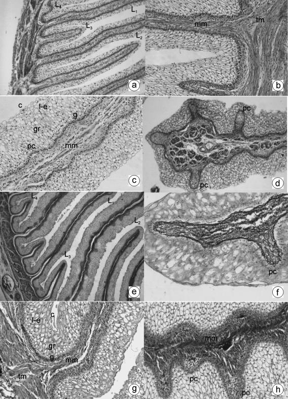
Fig. 2.
a Photomicrograph of a transverse direction section of the omasal wall at 21 cm CRL, 142 days. Omasal wall with the four laminar generations: primary (L1), secondary (L2), tertiary (L3) and quaternary (L4). H-E, ×120. b Photomicrograph of a transverse direction section of the omasal wall at 21 cm CRL, 142 days. Internal fascicule of the tunica muscularis (tm) filling the width of the smaller laminae and forming the muscularis mucosae (mm). MT, ×350. c Photomicrograph of a transverse direction section of the omasal wall at 33 cm CRL, 191 days. Primary lamina with stratified epithelium: stratum germinativum (g), granulosum (gr), lucidum-spinosum (l-e) and corneum (c). Corneum papillae (pc) and muscularis mucosae (mm) can also be seen (pc). H-E, ×250. d Photomicrograph of a transverse direction section of the omasal wall at 33 cm CRL, 191 days. Presence of corneum papillae (pc) in the secondary smaller laminae. H-E, ×180. e Photomicrograph of a transverse direction section of the omasal wall at 36 cm CRL, 205 days. Omasal mucosa with the four sizes of smaller laminae: primary (L1), secondary (L2), tertiary (L3) and quaternary (L4). H-E, ×180. f Photomicrograph of a transverse direction section of the omasal wall at 36 cm CRL, 205 days. Presence of reticulin fibres in the interior of the corneum papillae (pc) of the primary smaller laminae. RG, ×350. g Photomicrograph of a transverse direction section of the omasal wall at 40 cm CRL, 235 days. Stratified epithelial layer: stratum germinativum (g), granulosum (gr), lucidum-spinosum (l-e) and corneum (c). Muscularis mucosae (mm) coming from the internal fascicule of the tunica muscularis (tm). VG, ×350. h Photomicrograph of a transverse direction section of the omasal wall at 40 cm CRL, 235 days. Abundant presence of corneum papillae (pc) in the omasal smaller laminae. A thick muscularis mucosae can also be observed (mm). TM, ×350.

