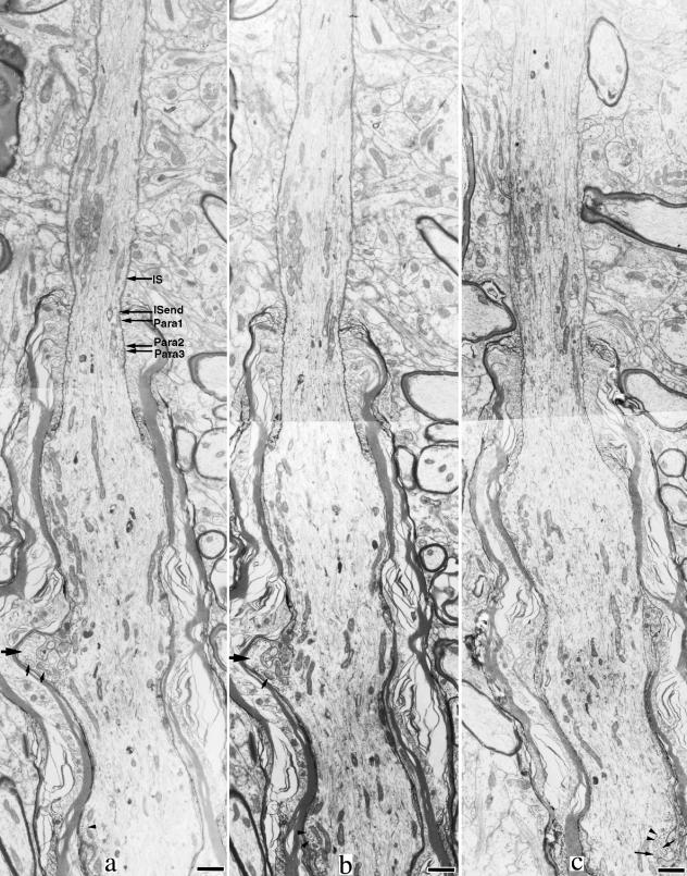
Fig. 2.
Serial electron micrographs of the distal part of the initial segment and the proximal part of the first internode. (a) Relative sites of five levels in Table 1 (IS, initial segment). Bar, 1.0 µm. The initial segment is the narrowest at the point where myelination starts. The paranode becomes thicker as the axon proceeds distally. The proximal part of the first internode shows a moderate enlargement. The internodal axon has a finger-like protrusion (large arrows). Near the axolemma, polyribosomes (arrowheads in panel c) and vesicular structures (small arrows) are observed.

