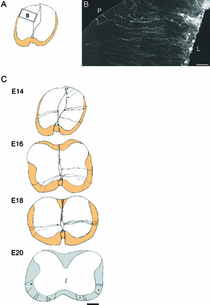
Fig. 1.
(A) line drawing with inset box indicates the positions in the spinal cord of the region shown in B. (B) Montage showing the distribution of DiI-labelled radial glia in the dorsal region of the spinal cord. Most cell bodies are closer to the lumenal than to the pial surface. Dotted line corresponds to the pial surface. L, luminal surface; P, pial surface. Bars, 40 µm. (C) Schematic representation of how the distribution of representative DiI-labelled radial glia changes from E14 to E20. Bar, 100 µm.

