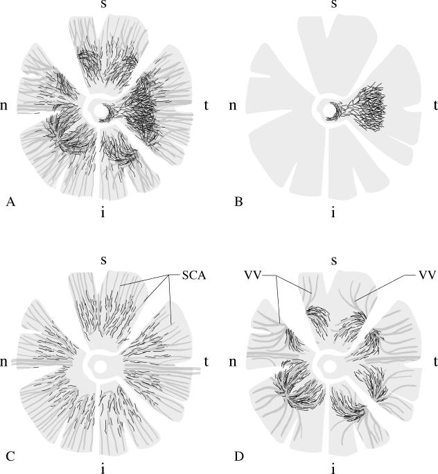
Fig. 1.
Schematic drawing of the distribution of non-vascular smooth muscle α-actin positive cells (NVSMC; black lines representing individual cells) in the different quadrants of a human choroid, using whole mount preparations. A: the complex arrangement of all NVSMC in the choroid. B: A plaque-like arrangement of NVSMC is present in the foveal region of the temporal quadrant, spreading up to the temporal rim of the optic nerve. C: In the posterior eye segment between the entry of the short posterior ciliary arteries (SCA) and the equator, the vessels are accompanied by NVSMC. NVSMC were no longer present in the peripheral choroid towards the ciliary body. D: Numerous NVSMC followed the course of the larger veins forming the vortex veins (VV) at the level of the equator bulbi. Again, the NVSMC were only present in the vessels draining the posterior choroid. The draining vessels from the ciliary body and the peripheral choroid were not paralleled by NVSMC. t = temporal, i = inferior, n = nasal, s = superior quadrant.

